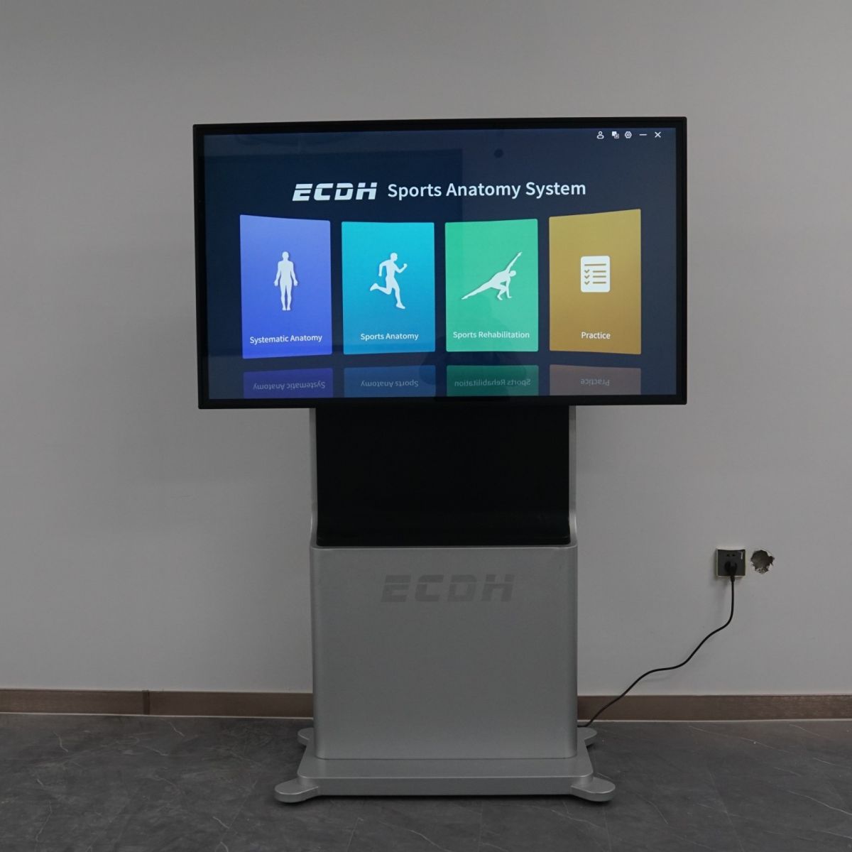
Digihuman Anatomy Teaching Blackboard
Equipment

Information
The Digihuman Virtual Anatomy Table contains 2 sets of complete human tomographic image data for male and female: 2110 layers for male, precision 0.1mm-1mm; 3640 layers for female, precision 0.1mm-0.5mm, which can display more than 6000 anatomical structures in 3D form, and is the only digital anatomy teaching product based on the reconstruction of complete tomographic data.
The system covers systematic anatomy, regional anatomy, tomographic images, clinical cases, etc. It is equipped with corresponding CT and MRI images (more than 1700) on the basis of tomographic specimen images, and has more than 130 anatomy teaching micro-class videos and a large number of digital practice questions.
Systematic Anatomy
The module contains 3D structures, obtained by 3D reconstruction of real human cross-sectional data. Their positions and shapes are consistent with the original data. These structures are divided into nine systems and can display the 3D morphology of more than 6000 anatomical structures.
Regional Anatomy
The module includes human peel and see-through features to demonstrate structures from superficial to deep, which makes it easy to build local hierarchical concepts and know the adjacent relationships of the structures even in the classroom. It also includes a large number of regional anatomy teaching videos to facilitate teaching and students’ self-study.
Sectional Anatomy
The module contains any part of the cross-sectional view. Using the highlighting function, the Chinese and English names of the cross-sectional structures can be quickly identified and their positions and shapes can be displayed in the 3D human body. Real specimens and images can be provided for students studying anatomy.
Anatomy Micro–course
The module include systematic anatomy micro course, regional anatomy micro-course and sectional anatomy micro course, and the number of courses is more than 130.
Autonomous Learning
